Science
AI Revolutionizes 3D Imaging of Biological Samples with New Algorithm
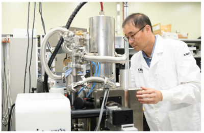
The Korea Research Institute of Standards and Science (KRISS), led by President Lee Ho Seong, has unveiled a groundbreaking artificial intelligence (AI)-based image segmentation algorithm. This innovative technology can quickly reconstruct three-dimensional (3D) structures from two-dimensional (2D) cross-sectional images of biological samples captured using a scanning electron microscope (SEM). This development has significant implications for various fields, including biology, materials science, and nanotechnology.
The algorithm enhances the efficiency and accuracy of 3D reconstruction, which has traditionally been a time-consuming and complex process. By leveraging advanced AI techniques, researchers can now obtain detailed 3D models from 2D images in a fraction of the time previously required. This capability not only streamlines research but also opens new avenues for understanding intricate biological processes and structures.
Transforming Research with AI
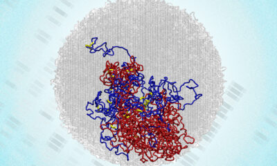
The implementation of this AI-driven approach allows scientists to visualize and analyze biological samples at a granular level. It is particularly beneficial for studies involving cellular structures and interactions. The ability to reconstruct 3D models enhances the understanding of how cells function and interact with their environment, which is crucial for advancements in medical research and development.
According to KRISS, the algorithm is designed to operate efficiently across a wide range of biological specimens, making it a versatile tool for researchers. The potential applications are vast, from pharmaceuticals to agricultural science, where understanding the microscopic world can lead to significant breakthroughs.
The development of this technology aligns with global trends in research, where AI is increasingly being integrated into scientific methodologies. The collaboration between AI and traditional imaging techniques promises to accelerate discoveries and improve the quality of research outputs.
Future Implications
As the demand for precise and rapid imaging techniques continues to grow, KRISS’s innovation positions it as a leader in the field of imaging technology. The integration of AI in microscopy not only enhances the accuracy of 3D reconstructions but also supports the ongoing shift towards more automated and sophisticated research methodologies.
The significance of this advancement cannot be overstated. It provides researchers with a powerful tool to explore and understand the complexities of biological systems in a way that was not previously possible. With this new algorithm, the future of microscopic imaging looks promising, paving the way for further exploration into the unseen world of cells and their functions.
In summary, KRISS’s AI-based image segmentation algorithm represents a significant leap forward in the field of microscopy. By transforming 2D images into detailed 3D models, it enhances our ability to study biological samples, which could lead to impactful scientific discoveries across various disciplines.

Woman Arrested for Threatening U-Haul Worker with Baton
Japan’s PM Takaichi Vows Urgent Fiscal Management Amid Rate Trends
Bitcoin Plummets Below $88K Amid Tether Concerns and Warnings

Tottenville Falls Short in PSAL 4A Title Game Against Erasmus

Shark Teeth Study Reveals Urgent Clues to Prevent Extinction

Church Members Rally Together After Thanksgiving Fire Devastation

U.S. Investment in Canadian Energy Firms Surges Despite Low Oil Prices

Nick Jonas Unveils New Solo Album ‘Sunday Best’ Set for February Release

Researchers Urge Label Changes for Low-Alcohol Drinks to Aid Pregnant Women

Urgent Update: Tom Aspinall’s Vision Deteriorates After UFC 321

MIT Scientists Uncover Surprising Genomic Loops During Cell Division

University of Hawaiʻi Joins $25.6M AI Project to Enhance Disaster Monitoring

AI Disruption: AWS Faces Threat as Startups Shift Cloud Focus

Time Crystals Revolutionize Quantum Computing Potential

Honeywell Forecasts Record Business Jet Deliveries Over Next Decade

Discover the Full Map of Pokémon Legends: Z-A’s Lumiose City

GOP Faces Backlash as Protests Surge Against Trump Policies

Parenthood Set to Depart Hulu: What Fans Need to Know
-
 Top Stories1 month ago
Urgent Update: Tom Aspinall’s Vision Deteriorates After UFC 321
-
 Health1 month ago
MIT Scientists Uncover Surprising Genomic Loops During Cell Division
-
Science4 weeks ago
University of Hawaiʻi Joins $25.6M AI Project to Enhance Disaster Monitoring
-
 Top Stories1 month ago
AI Disruption: AWS Faces Threat as Startups Shift Cloud Focus
-
 Science2 months ago
Time Crystals Revolutionize Quantum Computing Potential
-
 World2 months ago
Honeywell Forecasts Record Business Jet Deliveries Over Next Decade
-
 Entertainment1 month ago
Discover the Full Map of Pokémon Legends: Z-A’s Lumiose City
-
 Top Stories2 months ago
GOP Faces Backlash as Protests Surge Against Trump Policies
-
 Entertainment2 months ago
Parenthood Set to Depart Hulu: What Fans Need to Know
-

 Politics2 months ago
Judge Signals Dismissal of Chelsea Housing Case Citing AI Flaws
-

 Sports2 months ago
Yoshinobu Yamamoto Shines in Game 2, Leading Dodgers to Victory
-

 Health2 months ago
Maine Insurers Cut Medicare Advantage Plans Amid Cost Pressures