Science
University of Tokyo Develops Advanced Microscope for Cellular Insights
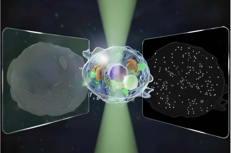
Researchers at the University of Tokyo have unveiled a groundbreaking microscope capable of detecting signals across an intensity range that is 14 times broader than that of traditional microscopes. This innovative device operates without the need for additional dyes, enabling label-free observations that are gentle on cells. This advancement holds significant promise for applications in pharmaceuticals and biotechnology, particularly in testing and quality control settings. The findings were detailed in the journal Nature Communications on November 14, 2025.
Microscopes have been instrumental in scientific progress since the 16th century. As demands for sensitivity and precision have evolved, researchers have faced challenges in balancing these needs with specialized techniques. For instance, quantitative phase microscopy (QPM) can identify structures larger than 100 nanometers but falls short when it comes to smaller entities. Conversely, interferometric scattering (iSCAT) microscopy excels at detecting single proteins, allowing researchers to track particles but lacks the comprehensive imaging capability of QPM.
Kohki Horie, one of the lead authors of the study, expressed a desire to explore dynamic processes within living cells using noninvasive techniques. To achieve this, the research team, which includes Horie, Keiichiro Toda, Takuma Nakamura, and Takuro Ideguchi, aimed to simultaneously measure both forward and backward light to capture a wide range of cellular sizes and movements within a single image.
To validate their concept, the researchers focused on observing the process of cell death. They successfully captured an image that incorporated data from both directions of light. “Our biggest challenge,” noted Toda, “was cleanly separating two kinds of signals from a single image while keeping noise low and avoiding mixing between them.” This approach allowed the team to quantify the motion of both micro-scale structures and nano-scale particles. By analyzing the forward and back-scattered light, they could estimate the size and refractive index of each particle.
Looking ahead, Toda remarked on the potential for future research, stating, “We plan to study even smaller particles, such as exosomes and viruses, and to estimate their size and refractive index in different samples. We also want to reveal how living cells move toward death by controlling their state and double-checking our results with other techniques.”
This pioneering work not only enhances current microscopy capabilities but also sets the stage for deeper insights into cellular dynamics and health-related research. As the field progresses, this technology could prove vital for understanding complex biological processes and improving diagnostic methods.
For further information, refer to the article titled “Bidirectional quantitative scattering microscopy” published in Nature Communications, DOI: 10.1038/s41467-025-65570-w.

Shark Teeth Study Reveals Urgent Clues to Prevent Extinction

Church Members Rally Together After Thanksgiving Fire Devastation

U.S. Investment in Canadian Energy Firms Surges Despite Low Oil Prices

Nick Jonas Unveils New Solo Album ‘Sunday Best’ Set for February Release

Researchers Urge Label Changes for Low-Alcohol Drinks to Aid Pregnant Women

Wedding Cookie Table Community Aims for World’s Largest Cookie Exchange

Mayor Appoints Alan Wong as New Supervisor for District 4

UK Report Reveals Growing Role of AI in University Research Assessments

Mayor Lurie Appoints Alan Wong as New District 4 Supervisor

Urgent Update: Tom Aspinall’s Vision Deteriorates After UFC 321

MIT Scientists Uncover Surprising Genomic Loops During Cell Division

University of Hawaiʻi Joins $25.6M AI Project to Enhance Disaster Monitoring

AI Disruption: AWS Faces Threat as Startups Shift Cloud Focus

Time Crystals Revolutionize Quantum Computing Potential

Honeywell Forecasts Record Business Jet Deliveries Over Next Decade

Discover the Full Map of Pokémon Legends: Z-A’s Lumiose City

GOP Faces Backlash as Protests Surge Against Trump Policies

Parenthood Set to Depart Hulu: What Fans Need to Know
-
 Top Stories1 month ago
Urgent Update: Tom Aspinall’s Vision Deteriorates After UFC 321
-
 Health1 month ago
MIT Scientists Uncover Surprising Genomic Loops During Cell Division
-
Science4 weeks ago
University of Hawaiʻi Joins $25.6M AI Project to Enhance Disaster Monitoring
-
 Top Stories1 month ago
AI Disruption: AWS Faces Threat as Startups Shift Cloud Focus
-
 Science2 months ago
Time Crystals Revolutionize Quantum Computing Potential
-
 World2 months ago
Honeywell Forecasts Record Business Jet Deliveries Over Next Decade
-
 Entertainment1 month ago
Discover the Full Map of Pokémon Legends: Z-A’s Lumiose City
-
 Top Stories2 months ago
GOP Faces Backlash as Protests Surge Against Trump Policies
-
 Entertainment2 months ago
Parenthood Set to Depart Hulu: What Fans Need to Know
-

 Politics2 months ago
Judge Signals Dismissal of Chelsea Housing Case Citing AI Flaws
-

 Sports2 months ago
Yoshinobu Yamamoto Shines in Game 2, Leading Dodgers to Victory
-

 Health2 months ago
Maine Insurers Cut Medicare Advantage Plans Amid Cost Pressures